Home
/ Tendon Diagram Of Wrist - Basic Hand And Wrist Anatomy Hand Institute Of Charleston / The carpal bones are arranged in 2 interrelated rows.
Tendon Diagram Of Wrist - Basic Hand And Wrist Anatomy Hand Institute Of Charleston / The carpal bones are arranged in 2 interrelated rows.
Tendon Diagram Of Wrist - Basic Hand And Wrist Anatomy Hand Institute Of Charleston / The carpal bones are arranged in 2 interrelated rows.. Its muscle belly is in the forearm and then travels along the inside of the forearm and crosses the wrist. It attaches to the base of the second and third hand bones. It also attaches to the trapezium, one of. Swelling of the tendons, and the tendon sheath, can cause pain and tenderness along the thumb side of the wrist. Wrist anatomy is the study of the bones, ligaments and other structures in the wrist.
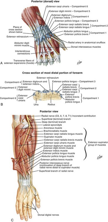
Superficial posterior muscles of the forearm posterior compartment muscles of the forearm. Posted on april 3, 2019april 3, 2019. The extensor tendon compartments of the wrist are six tunnels which transmit the long extensor tendons from the forearm into the hand. Swelling of the tendons, and the tendon sheath, can cause pain and tenderness along the thumb side of the wrist. A complete tear and a partial tear.
Wrist And Hand Clinical Gate from clinicalgate.com Gastrocnemius gastrocnemius muscle, large posterior muscle of the calf of the leg. Labeled body muscle diagram simple labeled muscle diagram. Related posts of wrist tendon anatomy diagrams shoulder bones anatomy diagram. The muscles and fasciae of the hand human anatomy / mri of finger ligaments radiology key. The wrist is a complex system of many small bones (known as the carpal bones) and ligaments. Wrist flexors the muscles attaching to the medial epicondyle and running down the front of the forearm that serve to flex the wrist and hand. They are located on the posterior aspect of the wrist. Forearm tendonitis is inflammation of the tendons of the forearm.
It also attaches to the one of the wrist bones, the trapezium.
They are located on the posterior aspect of the wrist. The wrist joint is a complex joint which connects the forearm to the hand, allowing a wide range of movement. The word tendinosis refers to a swelling of the tendons. The muscles and fasciae of the hand human anatomy / mri of finger ligaments radiology key. If any wrist tendonitis exercises cause you pain, stop immediately. 19 photos of the wrist tendon anatomy diagrams.extensor tendon compartments of the wrist are anatomical tunnels on the back of the wrist that contain tendons of muscles that extend (as opposed to flex) the wrist and the digits. Gastrocnemius gastrocnemius muscle, large posterior muscle of the calf of the leg. Anatomy diagrams of shoulder, arm, elbow, forearm, wrist and hand. Tendons are fibrous cords, similar to a rope, and are made of collagen. It also attaches to the trapezium, one of. Tendons are tissues that attach our muscles to our bones. Related photos for anatomy of the hand and wrist with tendons wrist tendons diagram preeminent wrist anatomy tendons at best. Ligaments connect one bone to another.
The carpal tunnel is a tube of nerves and tendons that passes through the wrist. Tendons are fibrous cords, similar to a rope, and are made of collagen. The tendon of extensor pollicis longus can be seen on the radial side of the wrist, at the base of the thumb where it forms the lower border of the 'anatomical snuffbox' a triangular shape between two tendons. Basic hand and wrist anatomy hand institute of charleston : This tendon is one of two tendons that bend the wrist.
Extensor And Flexor Tendon Injuries In The Hand Wrist And Foot Veterian Key from veteriankey.com Shoulder bones anatomy diagram 9 photos of the shoulder bones anatomy diagram acromion anatomy, ankle bones anatomy, bones of the clavicle, elbow bones anatomy, hip bones anatomy, shoulder muscles anatomy, shoulder pain anatomy, hand, acromion anatomy, ankle bones anatomy, bones of the clavicle, elbow bones. Evinrude 9.9 fuel pump diagram. Those that cross the palm side of the wrist are the flexor tendons. Forearm tendonitis is inflammation of the tendons of the forearm. Gastrocnemius gastrocnemius muscle, large posterior muscle of the calf of the leg. There are 6 tendons that help move your wrist. Both are made of collagen. These tendon sheaths allow the tendons to glide smoothly as the diagnosis of wrist tendonitis is made by looking for the characteristic signs of this condition.
Tendons are fibrous cords, similar to a rope, and are made of collagen.
You may be able to treat forearm tendonitis with rest and. Anatomy of the hand and wrist: Shoulder bones anatomy diagram 9 photos of the shoulder bones anatomy diagram acromion anatomy, ankle bones anatomy, bones of the clavicle, elbow bones anatomy, hip bones anatomy, shoulder muscles anatomy, shoulder pain anatomy, hand, acromion anatomy, ankle bones anatomy, bones of the clavicle, elbow bones. Related posts of wrist tendon anatomy diagrams shoulder bones anatomy diagram. Tendons are tissues that attach our muscles to our bones. The wrist links the hand to the forearm. Tendons to attach the muscles to the bones. Icing wrist tendonitis can help to cool inflammation and stimulates blood flow to the area of tendonitis. Tendon diagram ankle tendon diagram 9 out of 10 based on 90 ratings. Anatomy diagrams of shoulder, arm, elbow, forearm, wrist and hand. The wrist joint is a complex joint which connects the forearm to the hand, allowing a wide range of movement. The tendons that control movement in your hands, wrists and fingers run through your forearm. It also attaches to the one of the wrist bones, the trapezium.
Forearm tendonitis is inflammation of the tendons of the forearm. Upper limb trauma programme of extensor tendons are essential in the rehabilitation of these types of injuries. The paper linked below describes usual treatment for. Wrist flexors the muscles attaching to the medial epicondyle and running down the front of the forearm that serve to flex the wrist and hand. They curl the fingers and thumb, and they bend the wrist tendon diagram.
Tendons Hand Therapy Medical Anatomy Muscle Anatomy from i.pinimg.com It also attaches to the one of the wrist bones, the trapezium. If any wrist tendonitis exercises cause you pain, stop immediately. These tendon sheaths allow the tendons to glide smoothly as the diagnosis of wrist tendonitis is made by looking for the characteristic signs of this condition. Tendon diagram of wrist / transverse ultrasound of the first extensor compartment. The carpal tunnel is a tube of nerves and tendons that passes through the wrist. Gastrocnemius gastrocnemius muscle, large posterior muscle of the calf of the leg. This tendon works with the ecrb and ecrl to straighten the wrist. They are located on the posterior aspect of the wrist.
A complete tear and a partial tear.
The muscles and fasciae of the hand human anatomy / mri of finger ligaments radiology key. 19 photos of the wrist tendon anatomy diagrams.extensor tendon compartments of the wrist are anatomical tunnels on the back of the wrist that contain tendons of muscles that extend (as opposed to flex) the wrist and the digits. Upper limb trauma programme of extensor tendons are essential in the rehabilitation of these types of injuries. The word tendinosis refers to a swelling of the tendons. If any wrist tendonitis exercises cause you pain, stop immediately. The wrist joint is a complex joint which connects the forearm to the hand, allowing a wide range of movement. Related posts of wrist tendon anatomy diagrams shoulder bones anatomy diagram. Those that cross the palm side of the wrist are the flexor tendons. Tendons to attach the muscles to the bones. This tendon is one of two tendons that bend the wrist. The carpal tunnel is a tube of nerves and tendons that passes through the wrist. Wrist flexors the muscles attaching to the medial epicondyle and running down the front of the forearm that serve to flex the wrist and hand. They act with the profundus tendons to flex the wrist and mcp and pip joints.
The muscles that give rise to these tendons originate in the forearm and elbow ()the extrinsic extensor tendons reach the hand and digits by passing through a fibroosseous tendon sheath (retinaculum) located at the dorsal surface of the wrist tendon diagram. Its muscle belly is in the forearm and then travels along the inside of the forearm and crosses the wrist.

